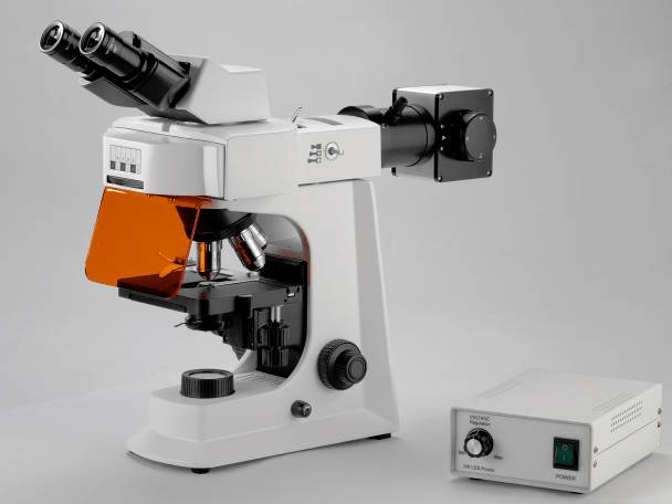
How Can a Fluorescence Microscope Be Combined With Other Imaging Techniques?

Fluorescence microscope excels at visualizing specific structures within a cell or sample thanks to fluorescent markers. However, it can sometimes lack detail on overall cell structure or lack the ability to see structures that don't have fluorescent tags. This is where combining fluorescence microscope with other imaging techniques becomes very powerful. Here are some ways fluorescence microscope can be integrated with other methods:
l Electron Microscope (EM): This powerful technique offers incredibly high resolution, allowing visualization of structures down to the atomic level. By combining fluorescence microscope for targeted labeling with EM for detailed structural analysis, researchers gain a much more comprehensive understanding of a sample. For instance, you could use fluorescence to identify specific proteins in a cell and then use EM to see their exact location and conformation within the cellular architecture.
l Brightfield/Light Microscope: This basic microscope technique provides an overall image of the sample with good contrast. Combining it with fluorescence allows researchers to visualize both the general cellular morphology and the specific fluorescently labeled structures within the same sample. This can be helpful for contextualizing the location of fluorescent markers within the larger cellular framework.
l Confocal Raman Microscope: This technique uses Raman spectroscopy to provide information on the chemical composition of a sample. Combining it with fluorescence microscopy allows for simultaneous analysis of both the spatial distribution of molecules (through fluorescence) and their chemical identity (through Raman spectroscopy). This can be particularly useful for studying the distribution of different biomolecules within a cell.
l Atomic Force Microscope (AFM): This technique provides information on the topography of a sample at the nanoscale. Combining it with fluorescence microscope allows researchers to correlate the spatial distribution of fluorescent markers with the surface features of the sample. For example, you could use fluorescence to identify specific proteins on the cell surface and then use AFM to measure the stiffness or topography of those regions.
These are just a few examples, and the specific combination of techniques will depend on the research question being addressed. The key takeaway is that combining fluorescence microscope with other imaging modalities offers a powerful approach to gaining a more comprehensive understanding of biological samples. You might also want to know how do I Prepare Samples for Fluorescence Microscope.
- Art
- Causes
- Crafts
- Dance
- Drinks
- Film
- Fitness
- Food
- Games
- Gardening
- Health
- Home
- Literature
- Music
- Networking
- Other
- Party
- Religion
- Shopping
- Sports
- Theater
- Wellness


